For advanced diagnostic services please visit our Specialty & Urgent Care Departments at 5 Cornerstone Square, Suite 203, Westford, MA 01886
Despite the very best preventive care, your pet can become sick. When this happens, we are here to provide advanced diagnostic services, including comprehensive internal medicine consultations, ultrasound, electrocardiograms, fluoroscopy and endoscopy that may not be available at your family veterinarian's hospital. Once completed, we will share the results with your family veterinarian to help formulate a treatment plan to return your companion to good health as soon as possible.
Internal Medicine Consultations
An important part of our services is the diagnosis and treatment of diseases that can affect your pet's internal organs and body systems. Our board-certified internal medicine specialists use advanced diagnostic technology to identify and effectively treat a range of complex internal medical issues.
During your initial visit, our veterinarian will talk with you, examine your pet and review your pet's medical history and any prior lab or X-ray results.
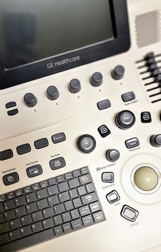
Ultrasound
Ultrasound employs sound waves in a painless, safe, non-invasive procedure to evaluate your pet's internal organs. It is often used as part of our comprehensive internal medicine consult. Ultrasound produces a real-time moving picture of your pet's organs, which allows us to visualize objects or possible abnormalities not detected by X-rays alone. We use ultrasounds to evaluate your pet's abdominal organs, and to identify masses or tumors. Ultrasound can also be used to help obtain samples for cytology or biopsy. In those cases we may recommend sedation or light anesthesia. A diagnosis as well as treatment options are often available immediately and can be explained during a consultation with one of our boarded internal medicine specialists.
An echocardiogram is an ultrasound of the heart. This test helps evaluate the size and function of the heart itself. Your family veterinarian may recommend an echocardiogram due to factors that may include:
- A new or worsening heart murmur
- Coughing
- Collapsing episode
- Breed predisposition
- Enlarged heart appearance on an x-ray, or other potential cardiac abnormalities.
This specialized test is available here and is performed by a board-certified cardiologist. A detailed report is written after the evaluation and forwarded to your family veterinarian to ensure continuity of care for your pet.
Endoscopy
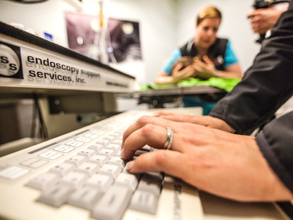
Endoscopy is a minimally invasive diagnostic procedure used to look at the interior surfaces of your pet's organs.
An endoscope is a medical device consisting of a long, thin, flexible (or rigid) tube which has a light and a video camera on the end that is inserted into your pet. Images of the inside of the patient's body are then viewed on a screen. It can often accurately diagnose issues that may be causing your pet's vomiting, diarrhea, weight loss, abdominal pain, abdominal distension, lack of appetite, nasal discharge, and certain respiratory problems.
In many cases it is considered a minimally invasive tool we can use to remove foreign objects, small polyps or tumors, and also use to collect biopsy samples. The benefits of endoscopy over exploratory surgery include no surgical incision, less time under anesthesia, less inflammation, less stress and discomfort and a quicker recovery time. Patients typically go home the same day as the procedure.
Our internal medicine department can perform a variety of endoscopic procedures, including:
- Upper and lower GI endoscopy- helpful in diagnosing potential health problems including certain forms of cancer or IBD.
- Removal of foreign objects from the stomach or esophagus.
- Cystoscopy
- Bronchoscopy and Bronchoalveolar Lavage
- Colonic/Esophageal stricture ballooning
- Rhinoscopy
- Otoscopy
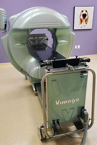
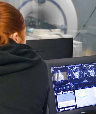
CT Scans
CT scanning, or computed tomography, creates high-resolution images of your pet. CT is one of the best diagnostic imaging tools available. The results are especially useful for detecting small masses and fully assessing damage in trauma situations, both of which may not be visible on conventional radiographs or ultrasound. The CT scanner is a ring-shaped apparatus that rotates around your pet. A computer builds an image of your pet, which can be manipulated in different ways, allowing for a comprehensive view of all internal organs. The detailed images produced by a CT scanner help us reach the best and quickest diagnosis possible.
One of the newest additions of advanced diagnostic tools is our Vimago CT machine. We are the only veterinary practice in the Merrimack Valley/Southern New Hampshire area with this equipment. This mobile robotic scanner can take nose to tail high definition, computed tomography-fluoroscopy-digital radiography pictures in a single session. The Vimago can accommodate the tiniest pets, as small as 2 ounces, or as large as 200 pounds. Most CT scans will require general anesthesia, but because of the speed of our machine, sedation can be used in certain cases. This revolutionary equipment is available for use throughout the hospital and has already helped save and improve the lives of our patients.
MRI
The electromagnet used in Magnetic Resonance Imaging (MRI) is a powerful tool used to examine soft tissues such as the brain, spinal cord, and nerves. The tube-shaped magnetic field and radio wave pulses can generate images of body structures.
An MRI can be used non-invasively to identify changes in delicate structures. Diseases such as stroke, bleeding, tumors, malformations, inflammation, infections, trauma and some degenerative conditions can be diagnosed on MRI.
Pets must go under general anesthesia during an MRI. Pre-anesthetic diagnostics such as blood panels, x-ray images, or ultrasound may be recommended in order to determine if your pet is a safe anesthetic candidate. A dedicated anesthesia technician will monitor your pet throughout the entire procedure.
Digital Radiographs
Radiographs, or X-rays, are one of the most common and useful diagnostic tools in medicine. After performing a thorough examination, diagnostic tests are sometimes necessary to identify the underlying cause of your companion's illness.
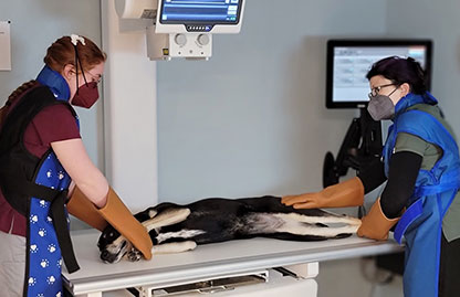
We use digital X-rays to examine your pet's bones, lungs, heart, abdomen, and other regions, to identify many medical and surgical conditions. There are many important advantages to digital versus conventional X-rays:
- They can be viewed immediately on our computer monitors by our veterinarians;
- The detailed images can be manipulated to clearly see your pet's bones and internal organs, leading to a faster, more accurate diagnosis;
- They take less time to process, which means your pet spends less time on the X-ray table, your pet's stress level is reduced and you receive the results quickly;
- No harsh chemicals are used to develop the images, reducing potential harm to our staff and the environment.
- Board-Certified Radiologist evaluation can be done in as little as 30 minutes.
- The images can be e-mailed or burned to a disc to share with your family veterinarian.
Advanced diagnostic capabilities are an extremely important part of emergency and referral veterinary medicine because certain conditions can be both urgent and complex. Our investment in digital X-ray technology reflects our commitment to offer your pet the best, most comprehensive veterinary care available.
Fluoroscopy
Fluoroscopy is a continuous X-ray that shows a real-time moving image of specific areas inside your pet's body. It allows our veterinarians to diagnose conditions that may have once required exploratory surgery, including:
- Tracheal Collapse
- Angiography - the imaging of blood flow through vessels using a special IV contrast dye.
- Swallowing disorders
With the use of this technology, we can offer your pet one of the most recent advancements in veterinary medicine.